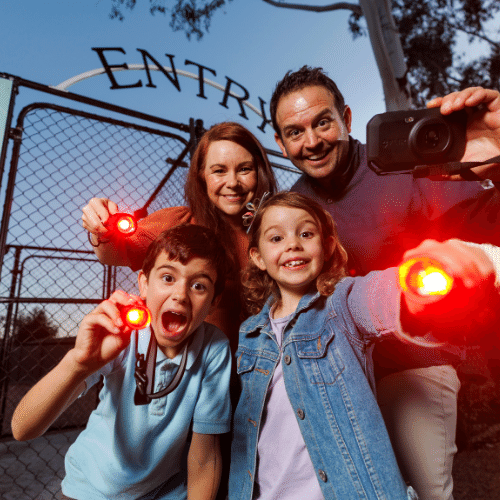
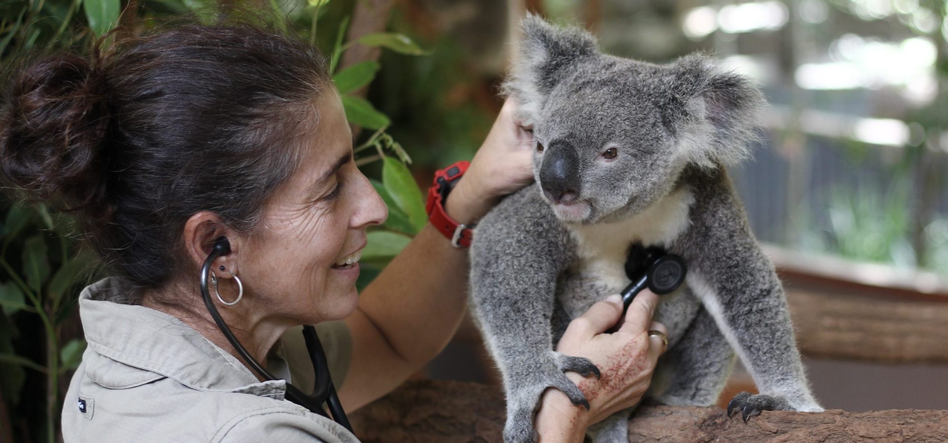
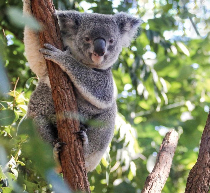

Lone Pine continues to research Australia's wildlife alone and in conjunction with scientists from Universities and the Government. Here are some of our published papers with their abstracts. For more information on this or other research, e-mail us at research@koala.net
![]()
Semen-induced luteal phase and identification of a LH surge in the koala (Phascolarctos cinereus)
D Johnston, P O'Callaghan, K Nilsson, G Tzipori and J D Curlewis
School of Animal Studies, The University of Queensland, 4072, Lone Pine Koala
Sanctuary, Jesmond Road, Fig Tree Pocket, 4069 and 2 School of Biomedical
Sciences, The University of Queensland, 4072, Australia
Correspondence should be addressed to 5 0 Johnston; Email:
s.johnston@uq.edu.au
The koala ovulates in response to mating. The purpose of this study was to document the LH surge induced by copulation and to investigate the potential roles of mechanical stimulation of the urogenital sinus and deposition of semen in induction of the luteal phase. In experiment 1, serial blood samples from four koalas that underwent normal mating showed elevated concentrations of LH approximately 24-32 h post-coitus. There was no corresponding elevation in LH in koalas (n = 4) that were exposed to the presence of a male but received no physical contact. In experiment 2, koalas on day 2 of oestrus were exposed to one of the following treatments (n = 9 per group): artificial insemination with 1 ml 0.9% sterile saline (control group), insemination with 1 ml koala semen, stimulation of the urogenital sinus with a purpose built glass rod (designed to mimic the action of the penis during natural mating) and urogenital stimulation with the glass rod followed by insemination of 1 ml koala semen. Confirmation of a luteal phase was based on evidence of a prolonged return to oestrus, parturition and/or elevated progesterone concentrations. Insemination of saline (0/9) and urogenital stimulation (0/9) failed to induce a luteal phase. Insemination of semen without glass rod stimulation resulted in a luteal phase in 4/9 koalas, three of which gave birth.
Insemination of semen in combination with urogenital stimulation produced a
luteal phase in 7/9 koalas, four of which gave birth. Semen had a significant
effect on induction of the koala luteal phase (P < 0.001) but glass rod
stimulation had no such effect (P = 0.335). It was concluded that semen must be
involved in the induction of a luteal phase in the koala. The results
presented in this study will serve to improve optimal timing and induction of
ovulation for artificial insemination in the koala. Reproduction (2004) 128
629-634
![]()
Natural and artificial methods for inducing the luteal phase in the koala (Phascolarctos cinereus)
S. D. Johnston, M. R. McGowan, P. O'Callaghan, R. Cox and V. Nicolson
School of Veterinary Science and Animal Production, The University of
Queensland, 4072, Australia; 2Lone Pine Koala Sanctuary, Jesmond Road, Fig Tree
Pocket, 4069, Australia; and 3Bioquest Ltd, North Ryde, 2113, Australia
.
An experiment was conducted in which female koalas were mated for different
durations of intromission and ejaculation to confirm that the luteal phase of
the oestrous cycle in koalas is induced by the physical act of mating. Results
showed that induction of a luteal phase in the koala usually required a complete
duration of penile thrusting behaviour from the male. It is proposed that
induction of a luteal phase in koalas may involve a copuloceptive reflex,
triggered by the thrusting of the male's penis into the female's urogenital
sinus. Although interrupted mating in koalas may be used to induce a luteal
phase in preparation for an artificial insemination programme, this study showed
that there is a 12.5% probability that pregnancy will result from semen
prematurely emitted by the teaser male. A dose of 250 iu hCG was administered
intramuscularly to eight oestrous females to determine whether it was possible
to induce a luteal phase artificially. In contrast to control females, which
received sterile saline injections, all females injected with hCG showed a
significant increase in progestogen concentration, above that of basal values,
indicating that a luteal phase had been induced successfully.
![]()
Studies of the oestrous cycle, oestrus and pregnancy in the koala (Phascolarctos cinereus
S. D. Johnston, M. R. McGowan, P. O'Callaghan, R. Cox and V. Nicolson
School of Veterinary Science and Animal Production, University of Queensland, 4072, Australia; Lone Pine Koala Sanctuary, Jesmond Road, Fig Tree Pocket, 4069, Australia; and 3Bioquest Ltd, North Ryde, 2113, Australia.
As an integral part of the development of an artificial insemination programme
in the captive koala, female reproductive physiology and behaviour were studied.
The oestrous cycle in non-mated and mated koalas was characterized by means of
behavioural oestrus, morphology of external genitalia and changes in the
peripheral plasma concentrations of oestradiol and progestogen. The mean (:!:
SEM) duration of the non-mated oestrous cycle and duration of oestrus in 12
koalas was 32.9 :!: 1.1 (n = 22) and 10.3 :!: 0.9 (n = 24) days, respectively.
Although the commencement of oestrous behaviour was associated with increasing
or high concentrations of oestradiol, there were no consistent changes in the
morphology or appearance of the clitoris, pericloacal region, pouch or mammary
teats that could be used to characterize the non-mated cycle. As progestogen
concentrations remained at basal values throughout the interoestrous period,
non-mated cycles were considered non-luteal and presumed anovulatory. After
mating of the 12 koalas, six females gave birth with a mean (:!: SEM) gestation
of 34.8 :!: 0.3 days, whereas the remaining six non-parturient females returned
to oestrus 49.5:!: 1.0 days later. After mating, oestrous behaviour ceased and
the progestogen profile showed a significant increase in both pregnant and
non-parturient females, indicating that a luteal phase had been induced by the
physical act of mating. Progestogen concentrations throughout the luteal phase
of the pregnant females were significantly higher than those of non-parturient
females. Parturition was associated with a decreasing concentration of
progestogen, which was increased above that of basal concentrations until 7 days
post partum.
![]()
Characterization of the Koala Biovar of Chlamydia pneumoniae at Four Gene Loci -ompAVD4, ompB, 16S rRNA, groESL Spacer Region
STACEY WARDROP, ANTHONY FOWLER, PAUL O'CALLAGHAN, PHILLIP GIFFARD, and PETER
TIMMS
Centre for Molecular Biotechnology, School of Life Sciences, Queensland
University of Technology, Brisbane, Australia Lone Pine Koala Sanctuary, Fig
Tree Pocket, Brisbane, Australia, 4001
Received November 2, 1998
Koalas are infected with two species of Chlamydia, C. pecorum and C. pneumoniae.
While it is known that significant genetic diversity occurs in the C. pecorum
strains infecting koalas, very little is known about the C. pneumoniae strains
that infect this host. In the current study, 10 isolates of koala C. pneumoniae
were analysed at four gene loci and found to be different to both the human and
horse C. pneumoniae strains at all loci (biovar differences ranging from 0.3%
at groESL up to 9.0% at ompAVD4). All koala biovar isolates studied were found
to be 100% identical at ompAVD4 (all 10 isolates) and at ompB (all three
isolates) genes. This lack of allelic polymorphisms at ompAVD4 has now been
observed for koala C. pneumoniae, human C. pneumoniae, guinea pig inclusion
conjunctivitis C. psittaci and feline conjunctivitis C. psittaci and may be
correlated to a lack of antibody response to the chlamydial major outer membrane
protein (MOMP) in these same strain/host combinations. This study also provides
the first documented case of natural C. pneumoniae infection causing a severe
and extended respiratory episode in a captive koala population. This captive
episode is in contrast to most free-range observations in which koala C.
pneumoniae is rarely documented as causing respiratory, ocular or urogenital
tract disease.
![]()
MEMOIRS OF THE QUEENSLAND MUSEUM
A RECORD OF COMMUNAL EGG LAYING IN THE SKINK CARLIA TETRADACTYLA (REPTILIA: SCINCIDAE). Memoirs of the Queensland Museum 33: 000-000. 1993:- During a visit to the Oakey area, southeast Queensland on 25 November 1992, a communal nest of 25 skink eggs was located. The site -a disused railway line was approximately)' 8km NW of Oakey township and consisted of a relatively continuous row of wooden sleepers. The nest was located beneath a single sleeper, which was lying on an approximate north-south axis, at its northern end. The whole sleeper was also angled slightly to the west. The eggs varied little in size. All were partially buried in the soil. The eggs were not measured at the time, but their size suggested they could belong to Carlia tetradactyla, a very common species at the site. Considering the paucity of records of communal nesting for Carlia, one egg was retained by one of us (RP) for artificial incubation to confirm its identification.
![]()
AN OBSERVATION OF INSECTIVOROUS FEEDING HABITS IN THE GREEN TREE SNAKE (DENDRELAPHIS PUNCTULATA)
Robert Porter, Lone Pine Koala Sanctuary, Jesmond Rd, Fig Tree Pocket QLD 4069
During a recent visit to Carnarvon Gorge, central Queensland, a brief
observation was made of an unusual feeding behaviour in the colubrid snake
Dendrelaphis punctulata. At around 1100 hours on a warm sunny day (temperature
approximately 28°C) in early March 1993, a green tree snake was seen amongst a
clump of sedge grass approximately 1 metre from the river's edge. The snake
appeared active and quite agitated, moving nervously in a short "start stop"
motion while holding an object in its jaws.
On closer inspection the item was seen to be some type of flying insect as a
pair of large, elliptical, transparent wings were projecting out of each side of
the mouth. The wings were consistent with a dragon -or damsel-fly, a lace wing
or a similar type of insect. The wings were seen to move up and down slightly,
though this appeared to be a consequence of the snake making a chewing motion
rather than movement from the insect. When approached closer the snake rapidly
fled into the clump of sedge and disappeared.
Many early authors listed insects as prey items for Australian snakes (e.g.
Kinghorn, 1929, Worrell, 1963, Gow, 1976). However, Shine (1991 a) states that
although insect fragments are often found in snake digestive tracts, in most
cases these were secondarily ingested from a vertebrate prey item. He does
indicate that occasional crickets have been found in some elapids and in his
examination of Australian colubrids (Shine, 1991 b) he lists 76 prey items from
58 D. punctulata, including one insect item, also a cricket. Considering the
huge number of Australian snake stomach contents examined by Shine over the last
15 years the occurrence of insects as prey items in Australian snakes (apart
from typhlopids) is evidently a very rare event.
It is possible that the insect observed being eaten by the green tree snake had
been captured by a frog or lizard which was then taken by the snake, both items
being swallowed consecutively. It seems likely however, that the frog or lizard
would have dropped the prey item when the snake struck. Even if this did occur,
the insect may have become contaminated by the lizard or frog's scent (in the
same fashion as keepers scent mice for lizard eating snakes) and the insect then
eaten independently by the snake. The most parsimonious explanation, however,
has to be that the snake captured and ate the insect itself, despite the rarity
of such events in Australian snakes. Though the importance of invertebrates in
most native snake diets is apparently negligible, this observation still raises
some interesting questions regarding the causes of such behaviour.
![]()
Effects of Acidic and Alkaline Treatments on Tannic Acid and its Binding Property to Protein
Ro Osawa and Terence P. Walsh
Veterinary Service and Research, Lone Pine Koala Sanctuary, Fig Tree Pocket, Brisbane, Queensland 4069, Australia, and Centre for Molecular Biotechnology, School of Life Science, Garden Point Campus, Queensland University of Technology, Brisbane, Queensland 4001, Australia
Effects of acidic and alkaline treatments on tannic acid (T A), one of the
hydrolyzable tannins, and its binding property to protein [bovine serum albumin
(BSA)] were investigated. Dissociation of TA-BSA complexes was apparent in both
highly acidic (pH <3) and alkaline (pH >7) treatments. T A dissociated from the
complexes and pure T A was not significantly hydrolyzed in highly acidic (pH
1.0-3.0) media and-retained its protein-binding property. TA did not seem to be
hydrolyzed even with a more acidic treatment (in 1 M HCI, pH <0.5), although its
protein-binding property was much reduced. The higher pH treatment (pH >6.-5)
facilitated the hydrolysis of T A, as judged by the release of its constituent
Gallic acid.
![]()
Metabolism of tannin-protein complex by facultatively anaerobic bacteria isolated from koala faeces
Ro Osawa, Terry P. Walsh & Steven J. Cork
Veterinary Service & Research, Lone Pine Koala Sanctuary, Fig Tree Pocket,
Brisbane, Queensland 4069, Australia,. Centre for Molecular Biotechnology,
School of Life Science, Queensland University of Technology, Faculty of Science,
Gardens Point Campus, Brisbane Queensland 4001, Australia, CSIRO Division of
Wildlife and Ecology, Lyneham, ACT2602, Australia (* Present address: Department
of Food Microbiology, School of Medicine, University of Tokushima, 18-15,
3-chome, Kuramotocho, Tokushima 770, Japan)
Received 10 June 1992; accepted in revised form 30 November 1992
The metabolic pathways involved in degradation of tannin-protein complex (T-PC)
were investigated in various facultatively anaerobic bacteria, with specific
reference to faecal isolates from the koala including T -PCdegrading
enterobacteria (T -PCDE), Streptococcus bovis, Klebsiellapneumoniae, and K.
oxytoca. It was demonstrated that ~-PCDE and S. bovis biotype I were capable of
degrading protein complexed with Gallo tannin (a hydrolyzable tannin), but not
that complexed with quebracho (a condensed tannin). Subsequent studies showed
that these strains metabolized Gallic acid to pyrogallol. Strains of Klebsiella
pneumoniae and K. oxytoca, which did not degrade T -PC, also metabolized Gallic
acid into pyrogallol. Pyrogallol was not degraded by any strains studied, but it
was not detected in fresh faeces of the koalas. The majority of strains isolated
from faeces could degrade phloroglucinol. Based on these findings, we propose
that members of the gut microflora of the koala cooperate in the degradation of
T -PC.
![]()
Microbiological Studies of the Intestinal Microflora of the Koala, Phascolarctos cinereus. II. Pap, a Special Maternal Faeces Consumed by Juvenile Koalas
R. Osawa, W. H. Blanshard and P. G. O'Callaghan
Veterinary Service and Research, Lone Pine Koala Sanctuary, Fig Tree Pocket, Qld 4069, Australia.
Quantitative and qualitative studies on the micro flora in the faeces of 10
female koalas, Phascolarctos cinereus, were conducted with specific reference to
'pap', a special maternal faeces consumed by the juvenile at about the time of
first emergence from the pouch. This specific coprophagy, called 'pap feeding',
occurred multiple times in all of the females examined. Pap was higher in water
content (81.8%) and pH (7.0) than normal faeces produced by the females before
and after pap feeding (54.6-56.4% and 5.5, respectively), suggesting that it is
derived directly from the contents of the caecum. Pap had higher (23-41-fold)
viable counts of tannin-protein-complex-degradi enterobacteria (T -PCDE) than
the normal faeces; in four of the females examined, viable T -PCDE were found in
pap but never in the normal faeces. The evidence indicates that pap feeding is
an essential physiological activity for the juvenile koala to prepare it for an
imminent dietary transition from maternal milk to
tannin-rich eucalypt leaves.
![]()
Visual Reading Method for Detection of Bacterial Tannase
R. OSAWA AND T. P. WALSH
Veterinary Service and Research" Lone Pine Koala Sanctuary" Fig Tree Pocket"
Brisbane" Queensland 4069" and Centre for Molecular Biotechnology" School of
Life Science" Queensland University of Technology"
Garden Point Campus" Brisbane" Queensland 4001, Australia
Received 19 October 1992/ Accepted 26 January 1993
Tannase activity of bacteria capable of degrading tannin-protein complexes was
determined by a newly developed visual reading method. The method is based on
two phenomena: (i) the ability of tannase to hydrolyze methyl gallate to release
free Gallic acid and (ii) the green to brown coloration of Gallic acid after
prolonged exposure to oxygen in an alkaline condition. The method has been
successfully used to detect the presence of tannase in the cultures of bacteria
capable of degrading tannin-protein complexes.
![]()
A Comparative Study of Macroscopic and Microscopic Dimensions of the Intestine in Five Macropods (Marsupialia: Macropodidae). I. Allometric Relationships
R. Osawa and P. F. Woodall
Department of Anatomy, University of Queensland, Brisbane, Qld 4072, Australia.
A Present address: Veterinary Service and Research, Lone Pine Koala Sanctuary,
Jesmond Road, Fig Tree Pocket, Qld 4069, Australia.
Abstract
Macroscopic and microscopic dimensions of the intestines in five macropod
species (the red kangaroo, Macropus rufus; the eastern grey kangaroo, M.
giganteus; the agile wallaby, M. agilis; the swamp wallaby, Wallabia bicolor;
and the red-necked pademelon, Thylogale thetis) were investigated
allometrically in relation to body mass.
In general, the length of the small intestine changed in an area: volume (A: V) compensating manner, but the circumference showed negative allometry such that the overall change in surface areas, both ground and mucosal (including the contribution of villi), were isometric but also included the coefficient derived from 'Kleiber's Law' (0' 75) in their 95 and 99% confidence limits, respectively.
Villous height and width generally showed no significant correlations with body size, but villous density was lower in large individuals.
The allometry coefficient for the length of the large intestine was generally
near the A: V compensating value (0' 5) in most intraspecific analyses but much
higher in the interspecific analysis, suggesting that some factor other than
body size might be important (possibly dietary fibre). Caecal length was
significantly correlated with body size only in two largest species (M. rufus
and M. giganteus) and the interspecific analysis gave a value near A: V
compensation).
![]()
Characterization of Gram-negative Anaerobic Strains, Isolated from Koala Feces, which Exhibit Satellite Growth and Pleomorphism
RO OSAWA, TOMOHIKO FUJISA WA, and TOMOT ARI MITSUOKA
Veterinary Service and Research, Lone Pine Koala Sanctuary, Fig Tree Pocket, Brisbane, Australia 4069, and Laboratory for Intestinal Flora, Frontier Research Program, Institute of Physical and 'Chemical Research (RIKEN), Wako-shi, Saitama 351-01, Japan
Received May 4, 1992
Three strains, which showed satellite growth around colonies of concomitant
isolates of Escherichia coli, were isolated from koala feces. Experimental
inoculation of these strains and E. coli strains on a plate medium anaerobic,
gram-negative, and highly pleomorphic rods. They were bile resistant and
asac-charolytic, and produced only a trace amount of propionic acid from peptone
yeast-extract Fildes-solution with glucose broth. Their growth was stimulated by
succinic acid but completely inhibited by fumaric acid. Subsequent biochemical
analysis revealed that the strains produced a large amount of propionic acid
from peptone yeast-extract (PY) broth supplemented with sodium succinate. A PY
broth agar plate, which contained 30% E. coli-metabolized PY broth, enhanced
growth of the strains, and it was found that this spent broth contained succinic
acid produced by the E. coli. The evidence suggests that the observed satellite
growth was due to succinic acid produced by E. coli strains.
![]()
Microflora of the Pouch of the Koala (Phascolarctos cinereus)
Ro Osawa, Wendy H. Blanshard and Paul G. O'Callaghan Veterinary Service and Research. Lone Pine Koala Sanctuary, .Jesmond Road, Fig Tree Pocket. Queensland 4069. Australia.
ABSTRACT: Microflora of the pouch epithelium of 17 female koalas (Phascolarctos cinereus) were examined in relation to their current reproductive status and recent reproductive history. No microbial growth was observed in pouch swabs from 13 of 17 (76%) koalas, including four females without young, seven with pouch young and two with back young (i.e. permanently emerged from the pouch). Growth of bacteria or yeasts was observed in pouch swabs from four koalas, each of which had experienced mortality of its pouch young during the current breeding season. Seven species of micro-organisms were isolated, including Pseudomonas aeruginosa, Serratia marcescens and Enterococcus faecalis. Based on the absence of microflora in the majority of females examined, we propose that the pouch epithelium normally provides a hostile environment for microbial colonization.
![]()
Tannin-Protein Complex-Degrading Enterobacteria Isolated from the Alimentary Tracts of Koalas and a Selective Medium for Their Enumeration
RO OSAWA
Veterinary Service and Research" Lone Pine Koala Sanctuary" Fig Tree Pocket"
Brisbane" Australia 4069
Received 23 October 1991/Accepted 7 March 1992
Tannin-protein complex (T -PC)-degrading enterobacteria (T -PCDE) were isolated
from the feces and from a layer of bacteria attached to the cecal wall of
koalas. The T -PCDE were facultatively anaerobic, gram-negative, pleomorphic,
nonmotile bacilli. The bacteria were also oxidase and catalase negative and
resistant to vancomycin, reduced nitrates to nitrites, and grew on MacConkey
agar. Growth on tannin-treated agar media showed a distinctive clear zone around
the colony. From these observations, a selective agar plate medium (vancomycin-
and tannin-treated Wilkins-Chalgren anaerobe. agar) was developed to enumerate
T-PCDE isolated from the feces of koalas. This medium was highly selective in
the enumeration of the fecal T -PCDE and inhibited the growth of concomitant T
-PC-degrading Streptococcus bovis. The T -PCDE were isolated from 10 of the 12
captive koalas studied; in 8 of these 10 koalas, the facultatively anaerobic
bacterial flora was dominated (more than 60%) by T-PCDE. Viable numbers of T-PCDE
were, in most of the animals, much larger (more than 100 times) than the numbers
of T -PC-degrading S. bovis, suggesting that T -PCDE played a more active role
in digesting T -PC in the alimentary tracts of koalas.
![]()
THE CURRENT STATUS OF THE KNOB-TAILED GECKOS (NEPHRURUS) IN CAPTIVITY
Robert Porter, Lone Pine Koala Sanctuary, Jesmond Road, Fig Tree Pocket, Brisbane, QLD 4069.
There are currently seven described species of Nephrurus, namely N. levis, N. laevissimus, N. asper, N. vertebralis, N. wheeleri, N. stellatus and N. deleani (Wilson and Knowles, 1988). They are all medium to large geckos with a very distinctive appearance, having a relatively large head with a short tail terminating in a small round knob. They are widespread over most of arid Australia, where they are endemic. At least one species, N. deleani, is very restricted in range (Wilson and Knowles, 1988). A detailed review of the biology and ecology of the genus can be found in Greer (1989).
This article was stimulated by discussions in meetings of the Reptile Taxon Advisory Group, the body responsible for the overall management of reptiles in Australasian zoos. Geckos of the genus Nephrurus were chosen for particular attention, among other taxa, due to the low level of knowledge of their reproductive biology and captive requirements, as well as their desirability for captive collections and their unique morphology. The group acknowledges that some of the animals on which this article is based are probably held illegally, but, while it in no way supports illegal take or trade, it places the wide dispersal of knowledge of our native herpetofauna as one of its highest priorities.
![]()
Phenotypic Characterization of CO2-Requiring Strains of Streptococcus bovis from koalas
R. OSAWA AND L. I. SLY
Veterinary Service and Research, Lone Pine Koala Sanctuary, Fig Tree Pocket,1
and Department of Microbiology, University of Queensland, St. Lucia,2 Brisbane,
Queensland, Australia
Received 16 April 1991/ Accepted 29 July 1991
We examined phenotypic characteristics of six mannitol-fermenting strains of Streptococcus bovis, including two unusual CO2-requiring strains isolated from koala feces. These strains did not grow in air, but grew in air supplemented with CO2 and under reduced oxygen conditions. All six strains had the same biochemical characteristics, except that the CO2-requiring strains did not produce p-N -acetylglucosaminidase.
Streptococcus bovis is commonly found in the normal gastrointestinal floras of many animals (2, 3, 8, 16) and also is often isolated in clinical situations, such as cases of human bacteremia (9, 10, 13), endocarditis (7, 17), and bovine mastitis and udder infections (5, 12). Although S. bovis is usually considered to be a facultative anaerobe (6), Latham et al. (11) reported that this species includes strains which are strictly anaerobic when they are first isolated. Recently, Osawa (14) and Osawa and Mitsuoka (15) investigated the phenotypic characteristics of facultatively anaerobic strains of S. bovis obtained from various sources. We isolated apparently anaerobic strains from the feces of two healthy koalas (Phascolarctos cine reus (Goldfuss), and in this study we characterized these strains.
Six strains of S. bovis, including two that are not able to grow in air, were obtained from our collections at the Lone Pine Koala Sanctuary (strains LPKS 1, LPKS 6, LPKS 31, and LPKS 32) and the Department of Microbiology, University of Queensland, Queensland, Brisbane, Australia (strains UQM 3549 and UQM 3552). The sources of the strains are indicated in Table 1. Cultures of these strains, which were stored as lyophiles, were revived and subcultured anaerobically at least three times on plates containing Columbia blood agar (Oxoid, Ltd., Basingstoke, Hampshire, United Kingdom) in an atmosphere enriched with 8 to 10% CO2 by using Bio-bags (Becton Dickinson and Co., Cockeysville, Md.) before use.
![]()
Serological assessment of chlamydial infection in the koala by a slide EIA technique
H UENO, S MIZUNO, I TAKASHIMA, R OSAWA, W BLANSHARD, P TIMMS, N WHITE, N HASHIMOTO
Summary: A rapid and simplified slide enzyme immunosorbent assay (EIA) was
developed for the diagnosis of chlamydial infection In the koala. HeLa 229 cells
Infected with koala strain Chlamydia psittaci were fixed on the surface of
multiwell slides and used as the antigen. The assay consisted of first reacting
koala antiserum with the fixed C psittaci antigen, followed by reaction with
biotinylated rabbit anti-koala IgG, ABC reagent and substrate. The chlamydial
EIA antibody titres obtained were compared with those of a complement fixation
(CF) test using koala strain C psittaci as antigen. Of 35 koala sera tested, 16
CF positive sera (~1: 8) also had a positive titre (~1: 200) In the slide EIA
test (sensitivity 93.8%, 15/16). Nineteen CF negative sera were also negative in
the slide ElA (specificity 100%, 19/19). Sixty-eight samples of koala blood were
collected by ear-prick using a sampling paper method and were assayed by both
tests. Sensitivity of the slide EIA was 100% (15/15) and specificity of the test
was 96.2% (51/53). To simplify the slide EIA for use as a practical screening
test, a 3-polnt serum dilution series (1:100, 1: 200, 1: 400) was used. This
3-polnt slide EIA was compared with the CF test using sheep strain chlamydial
antigen. Thirty-nine sera were assayed by both tests. The sensitivity of the
3-polnt method was 85.7% (6n) and the specificity was 71.9% (23/32) as compared
with the sheep antigen CF test. Of the 10 koalas with discrepant results, 8
(slide EIA positive but sheep antigen CF negative) were either chlamydia
positive in cell culture or had evidence of clinical symptoms. This suggests a
significant superiority of the slide EIA for detecting Infected animals as
compared with the sheep antigen CF test. The 3-polnt slide EIA was also applied
to cephalic vein blood collected on sampling paper and good agreement was
obtained In 25 of 26 (96.1 %) samples when compared with those of serum samples.
These results show that the slide EIA Is at least equal to the koala antigen CF
test, and significantly more sensitive than the sheep antigen CF test for the
serological assessment of chlamydial infections in koalas. Aust Vet J 68: 393
E96.
![]()
UNUSUAL BASKING BEHAVIOUR IN CAPTIVE BEARDED DRAGONS (POGONA BARBATA)
Robert Porter Lone Pine Koala Sanctuary, Jesmond Road, Fig Tree Pocket, QLD, 4069.
Two fourteen month old, captive hatched Bearded Dragons (Pogona barbata) are currently maintained in an inside enclosure measuring 2400mm x 900mm x 600m high with two adult Shingleback skinks (Tiliqua rugosa). The substrate consists of a layer of dry mulched eucalyptus branches and the inhabitants are supplied with raised basking sites in the form of rocks and branches. Light and heat are provided by two 150 watt white spotlights plus a single 4-foot fluorescent fitting containing an F40T9BL "black light" tube. All are suspended 500mm above the substrate, although the lizards can move closer by climbing onto the basking sites. The enclosure is housed in an air conditioned room which produces a heat gradient of 36° 37°C directly beneath the spotlights to around 23° -24°C in the coolest corner. The lights are controlled automatically by an electronic timer to simulate the local natural photoperiod. These lizards have been maintained inside with no access to direct sunlight for at least ten months with no apparent health problems.
For a period of three days in mid January 1991 all four lizards were removed to a 900mm x 300mm x 400mm glass tank while their permanent enclosure was thoroughly cleaned. A wooden lid covered with insect mesh and 12mm weld mesh for ventilation was fitted to the top. An incandescent light fitting was situated in the middle of this lid and contained a 75 watt white bulb. Rocks were placed directly beneath the bulb allowing the lizards to bask within 200mm of the light. Unfortunately no temperatures were taken in the enclosure but an estimation would be about 25° -28°C.
![]()
Application of a blood sampling paper method for complement fixation test detection of anti-chlamydial antibody in koalas (Phascolarctos cinereus)
R. OSAWA, F. N. CARRICK, N. HASHIMOT0, I. TAKASHIMA and T. TAKAHASI.
Blood samples obtained from 23 koalas were examined for detection of anti-chlamydia CF antibody by two different methods: (1) a conventional CF test on serum; (2) a new CF test on whole blood absorbed in a Blood Sampling Paper. The results obtained from these methods showed good correlation, with consistency in 22 out of 23 samples. This new CF test does not require a large amount of blood, and this may be obtained by pricking the ear vein rather than the more invasive cephalic vein puncture conventionally used. Thus this method greatly facilitates and simplifies the procedure of assessing anti-chlamydia CF antibody status of koalas, which are easily "stressed'' by handling, and seems to provide a more robust technique for monitoring koala populations in the field.
Introduction
INFECTIONS with bacteria belonging to the genus Chlamydia are widespread in the
animal kingdom (Storz 1971). One of the most common methods used to screen for
infection by these organisms is an assay which detects complement fixing (CF)
antibodies to Chlamydia sp. in the blood. Conventional CF tests require 0.2 ml'
of serum which would normally be derived from 1 ml of whole blood drawn by
venipuncture. This technique is frequently used to screen for Chlamydia psittaci
infections in captive colonies of koalas.
The Nobuto Blood Sampling Paper (Toyo Roshi Kaisha Ltd, Tokyo) has been
developed as an alternative procedure for the collection, transportation and
storage of blood for serological examination, and has potential for use by
personnel with no experience in collecting blood by venipuncture. This technique
has been used successfully for a wide variety of serological examinations in the
microbiological field and is particularly convenient for continuous testing of
newborn and small animals, and in surveys. Examples of prior application of the
device include studies of toxoplasmosis (Nobuto 1965a), Japanese encephalitis,
Newcastle disease, mycoplasmosis and canine distemper (Nobuto 1965b).
The objectives of this study were to evaluate whether samples of whole blood collected from koalas by this method are suitable for use in a CF test, and to compare the performance of the CF test on these samples with the conventional method which utilizes blood serum.
![]()
Feeding Strategies of the Swamp Wallaby, Wallabia bicolor, on North Stradbroke Island, Queensland. I: Composition of Diets
Ro Osawa
Department of Anatomy, University of Queensland, St Lucia, Qld 4072; present address: Division of Veterinary Service and Research, Lone Pine Koala Sanctuary, Jesmond Road, Fig Tree Pocket, Qld 4069, Australia.
Abstract
Species composition of plants present in the stomach contents of 34 road-killed
swamp wallabies, W. bicolor, on North Stradbroke Island, south-east Queensland,
was investigated, and related to seasonal variations in nitrogen and fibre
contents of plants in the study area. W. bicolor consumed a wide range of plant
categories including shrubs, forbs, grasses, ferns and fungi. Among these,
grasses, followed by shrubs, were the major component of the stomach contents.
These dietary plants had higher nitrogen contents than those non-dietary plants
of the corresponding plant category, which were present in the study site but
never found in the stomach contents. Individual grass species of high occurrence
in the stomach contents were molasses grass, Melinus minutiflora, green couch,
Cynodon dactylon and sedges, Cyperus spp. Although more fibrous, these species
had similar or relatively high nitrogen levels compared to native shrubs, except
during summer. This suggests that W. bicolor selected dietary plants of high
nitrogen content rather than of low fibre content.
![]()
Feeding Strategies of the Swamp Wallaby, Wallabia
bicolor, on North Stradbroke Island, Queensland.
II: Effects of Seasonal Changes in Diet Quality
on Intestinal Morphology
Ro Osawa and Peter F. WoodaII
Department of Anatomy, University of Queensland, St Lucia, Qld 4072, Australia.
A Present address: Division of Veterinary Research, Lone Pine Koala Sanctuary,
Jesmond Road, Fig Tree Pocket, Qld 4068, Australia.
Abstract
The effect of seasonal changes in diet quality on the alimentary tract of the
swamp wallaby, W.bicolor, on North Stradbroke Island was investigated by
examining 25 road-killed animals. To allow for variation in body weight,
allometric analyses of both macroscopic and microscopic dimensions of the
intestine were used and differences were tested by analysis of covariance. W.
bicolor on a high fibre diet in spring and autumn had significantly shorter
villi compared to those on a low fibre diet in winter and summer, suggesting
that the intestinal villi of W. bicolor are highly labile and respond rapidly to
changes in diet quality.
![]()
Formation of a Clear Zone on Tannin-Treated Brain Heart Infusion Agar by a Streptococcus sp. Isolated from Feces of Koalas RO OSAWA
Veterinary Service and Research, Lone Pine Koala Sanctuary, Fig Tree Pocket, Brisbane, Queensland 4069, Australia
Received 5 October 1989/ Accepted 7 December 1989
Gram-positive cocci, isolated from the feces of koalas and identified as Streptococcus bovis biotype I, formed a distinct clear zone on tannin-treated brain heart infusion agar, suggesting that this isolate has the unique characteristic of degrading the tannin-protein complex.
The koala, Phascolarctos cinereus (Goldfuss), is an arboreal marsupial inhabiting the forests of eastern Australia. It is entirely folivorous, feeding almost exclusively on the foliage of Eucalyptus spp. (5, 6), which are known to have high concentration of tannins (3, 11). Tannins are a diverse group of soluble phenolic compounds that form chemical complexes with proteins. These complexes are considered to be resistant to degradation (i.e., by protease) within the guts of mammals, thereby interfering with digestion and the utilization of dietary protein (8, 13, 16, 18). Cork et al. (2) found a low tannin level in koala feces relative to that in the diet and- suggested that the tannin fraction of the total phenolic compounds was degraded to a considerable extent in the alimentary tracts of koalas. Investigation of the functions of the ceca of the koala (12) and the ring tail possum, Pseudocherius peregrinus, another marsupial feeding on eucalyptus diets (A. Lomdahl, Hons. thesis, Monash University, Clayton, 1983), led to the claim that bacterial degradation of tannin-protein complexes in the cecum may make protein available to these animals.
Fresh fecal pellets were collected directly from the anogenital orifice of 12
captive koalas (nine adult males and three adult females) kept at the Lone Pine
Koala Sanctuary. The animals were provided daily with fresh eucalyptus leaves ad
libitum. The major species of eucalyptus leaves fed to the animals included
forest red gum (Eucalyptus tereticornis), tallow wood (E. microcorys), and.
grey gum (E. punctata). Fecal pellets collected from each animal were weighed
aseptically, and approximately 1 to 2 g (wet weight) was taken into a tube
containing 20 ml of sterile 0.25-strength Ringer solution (Oxoid Ltd.,
Basingstoke, Hampshire, United Kingdom) and thoroughly mixed with a homogenizer
and a Vortex test-tube mixer.
![]()
Growth and development of the koala from birth to weaning
WENDY H. BLANSHARD
Changes in the appearance, dentition and behaviour of juvenile koalas during the first year of life are described. A number of these changes are discussed in relation to the normal timetable of physical and behavioural development of koalas reared naturally by their mothers in zoos and sanctuaries.
INTRODUCTION
IN common with other marsupials, koalas (Phascolarctos cinereus) are born in a
very undeveloped state, weighing less than one gram. Although the respiratory,
digestive and urinary tracts are functional and the forelimbs are already
equipped with claws, the hind limbs are little more than buds (Cork 1987a; Lee
and Martin 1988). During the first year of life most of the physical attributes
of an adult koala develop, and the young koala becomes physically and
physiologically independent of the mother.
Little published information is available on the growth and development of juvenile koalas. Eberhard (1972) examined a number of pouch young of the Victorian race (P. cinereus victor) occurring on Kangaroo Island, South Australia, and produced a schedule of first appearance of some external characteristics. As the ages of these animals were unknown, his descriptions were tabulated against body weight and various anatomical measurements. Lee and Martin (1988) gave an outline of the development of P. cinereus victor at intervals between birth and independence, including general information on body weight, head length, physical appearance, dentition and behaviour.
Only Smith (1975) and Thompson (1987) have documented the exact ages of individual koalas when describing aspects of their development. Both authors studied captive-bred animals of the Queensland race (P. cinereus adustus), and recorded stages in their behavioural and dietary transition from life inside to life outside the pouch.
In a search for body traits which may be useful in predicting the age of koalas,
Eberhard (1972) found that head length, elbow-wrist length and ankle-knee length
were linearly related to each other over a wide range of ages. Both Eberhard
(1972) and Martin (1983) plotted the relationships between head length and
estimated age for different populations of Victorian koalas. The youngest age
represented on any of these plots was 200 days (Martin 1983).
Over the past three years, the author has accepted 21 juvenile Queensland koalas
(P. cinereus adustus) for hand-raising. These comprised nine unfurred and four
partially furred pouch-young, and eight fully furred back-young. A quantitative
method of estimating the ages of the hand-raised koalas was devised, so that
their growth and development relative to one another could be meaningfully
assessed. This paper documents some of the physical and behavioural changes
which occur during the first 12 months of life of the koala, encompassing the
period from birth to weaning.
![]()
Selective Medium for Enumeration of Tannin-Protein Complex Degrading-Streptococcus spp. in Feces of Koalas
RO OSAWA AND TOMOTARI MITSUOKA
Received 15 June 1990/ Accepted 6 September 1990
A selective agar plate medium (tannin-treated brain heart infusion agar supplemented with colistin-oxolinic acid) was developed to enumerate tannin-protein complex-degrading Streptococcus bovis in the feces of koalas. This medium was successfully used to enumerate strains from fecal samples but failed to enumerate those from pure cultures.
Streptococcus bovis is a common isolate from the alimentary tracts of ruminants (4, 6, 12, 17) and nonruminants (1, 15). Strains of bovine origin are frequently found to belong to biotype II (mannitol nonfermenting), whereas strains of human origin, which are associated with human systemic infections, have more often been found to belong to biotype I (mannitol fermenting) (5, 7, 11, 16). Recently, Osawa (9) reported that S. bovis biotype I isolated from koala (Phascolarctos cinereus) feces had the unique characteristic of growing on tannin-treated brain heart infusion agar (TTBHIA) and forming a clear zone around the colony. In the same work, a strain of S. bovis biotype II used for comparison grew on T-TBHIA plates but did not form a clear zone. Strains of S. faecalis (Enterococcusfaecalis) and S. faecium (Enterococcus faecium) did not grow on this medium. On the basis of these findings, Osawa (9) suggested that S. bovis biotype I is capable of degrading the tannin-protein complex, thereby helping koalas obtain dietary protein from tanninrich eucalypts.
This report describes a selective medium used to enumerate the strains degrading the tannin-protein complexes. Such a medium would allow us to monitor the tannin-protein complex -degrading activity of the gastrointestinal microflora in koalas, whose diet consists almost exclusively of tanninrich eucalyptus leaves (3).
The strains of S. bovis and other streptococcal and enterobacterial strains used for comparisons in this study are listed in Table 1. Cultures of the strains were maintained on BHIA (Oxoid Ltd., Basingstoke, Hampshire, United Kingdom) supplemented with 0.5% yeast extract (Oxoid). Physiological characteristics of the streptococci were determined by the API 20 Strep system (API System, Montalieu, Vercieu, France) to ensure their identity immediately before inoculation onto test plates. The identities of the enterobacterial strains were checked by standard methods.
![]()
Use of a dietary supplement in koalas during systemic antibiotic treatment of chlamydial infection
Department of Zoology, University of Queensland, St Lucia, Queensland 4067
R OSAWA FN CARRICK
Infection with Chlamydia psittaci in the koala can cause blindness, pneumonia, infertility and urinary tract diseases and has been described by many workers (Cockram and Jackson 1974; Dickens 1976; Cockram 1978; Brown eta11980; Brown and Grice 1984). A survey of both captive and wild koalas throughout eastern Australia from 1979 to 1985 indicated that a large proportion of koalas sampled was affected by C psittaci (Brown and Carrick 1985).
Ad hoc attempts to treat chlamydial infections in koalas with antibiotics usually failed, although Carrick and Wood (1986) found that administration of dietary supplements enabled the majority of koalas treated with oxytetracycline by injection to survive.
Nevertheless, Brown (1987) reported that systemic antibiotic therapy for chlamydial infections in koalas, using either erythromycin or oxytetracylcine, was invariably fatal. The koalas showed rapid loss of body weight and died, although there was a marked reduction in signs of chlamydiosis during the course of the treatments.
As suggested by Carrick and Wood (1986), supplementary feeding using soya-based
infant feeding preparation has become a common management practice for captive
koalas in many zoos, including Lone Pine Koala Sanctuary in Australia, and
Saitama Children's Zoo and Awaji Farm Park in Japan. Handasysde et al (1988)
have also since recommended such supplementary feeding for captive koalas
showing consistent loss of weight. We have conducted a controlled experiment to
evaluate the efficacy of such dietary supplementation during systemic
antibiotic' therapy in koalas.
![]()
Road-kills of the Swamp Wallaby, Wallabia bicolor, on North Stradbroke Island, South-east Queensland
Ro Osawa
Department of Anatomy, University of Queensland, St Lucia, Qld 4067., present address: Division of Veterinary Service and Research, Lone Pine Koala Sanctuary, Jesmond Road, Fig Tree Pocket, Qld 4069.
Abstract
A total of 127 road-kills of the swamp wallaby, Wallabia bicolor, on North
Stradbroke Island in south-east Queensland was recorded between May 1981 and
April 1982. The majority of kills were of adults. Road-kills occurred mainly at
night, suggesting that W. bicolor is nocturnal, and were significantly
correlated with the number of vehicles brought onto the island by ferries.
Faecal pellet counts were made in classified habitats along the road in order to
determine the relationship between the population density and road-kills of W.
bicolor. Road-kills were significantly correlated with the faecal pellet counts
in the Disturbed Ares (narrow, artificially cleared areas adjacent to the road)
but not with the counts in the Native Area (i.e. native forest). This indicates
that the wallabies were attracted to the disturbed area and became more
susceptible to being killed by vehicles. There was no significant variation in
the number of road-kills between lunar phases.
![]()
FAECAL MICRO FLORA OF CAPTIVE KOALAS, PHASCOLARCTOS CINEREUS (MARSUPIALIA: PHASCOLARCTIDAE)
Ro OSAWA AND TOMOTARI MITSUOKA
Osawa, R. and Mitsuoka, T. 1990. Faecal microflora of captive koalas,
Phascolarctos cinereus
(Marsupialia: Phascolarctidae). Australian Mammalogy 13: 141-147.
The faecal microflora of four Phascolarctos cinereus kept at the Saitama Children's Zoo in Japan was investigated using 10 selective and 4 nonselective agar media. Total viable counts (in log form) of faecal bacteria (8.2 g of wet faeces) were significantly lower than the direct microscopic counts (11.2 g of wet faeces). Four groups of obligate anaerobes (bacteroidaceae, peptococcaceae, bifidobacteria, and clostridia) and three groups of facultative anaerobes (enterobacteria, streptococci and lactobacilli) were isolated. Dominant components of the flora were obligate anaerobes, in particular bacteroidaceae and peptococcaceae.
R. Osawa, Department of Zoology, University of Queensland, St Lucia, Queensland 4067 Australia. Present address: Veterinary Service and Research, Lone Pine Koala Sanctuary, Jesmond Road, Fig Tree Pocket, Queensland 4069 Australia. 7: Mitsuoka, International Frontiel; Research System, RIKEN, Wako, Saitama 351. Department of Biomedical Science, Faculty of Agriculture, The University of Tokyo, Bunkyo, Tokyo, 113 Japan. Manuscript received 21 September 1989.
KOALAS (Phascolarctos cinereus) are arboreal marsupials living mainly in the
forests of eastern Australia. The animal feeds entirely on a folivorous diet,
consisting almost exclusively of foliage of trees of the genus Eucalyptus (Eberhard,
McNamara, Pearse and Southwell 1975, Eberhard 1978).
McKenzie (1978) observed that the luminal surface of the caecum was completely
covered by a tangled mat of bacteria including gram-positive and gram-negative
rods, cocci and actinomycete-like organisms. He also suggested that the caecum
of P. cinereus was a major site of microbial fermentation of ingesta. However,
Cork, Hume and Dawson (1983) reported that when P. cinereus were fed on a diet
of the grey gum (Eucalyptus punctata) only about 9 070 of total energy intake
was derived from volatile fatty acid production; thus suggesting that the major
role of the hindgut micro flora of P. cinereus may not be fermentation of plant
cell wall cellulose.
In the present study, the composition of faecal microflora of captive P. cinereus kept at a Japanese Zoo was investigated in order to provide information on the ecology of the gut microflora. Since information on the composition of faecal flora of other animals was provided by Mitsuoka and Kaneuchi (1977), the methods of culturing, isolating, identification, and enumeration used by these workers were used in the present study for quantitative comparison between P. cinereus and those animals.
![]()
New Zealand Geckos
Their Natural History and Captive Maintenance
Robert Porter
c/o Lone Pine Koala Sanctuary
Brisbane, Queensland 4069 –Australia
Jesmond Road, Fig Tree Pocket
Isolated from other landmasses for millions of years, New Zealand's terrestrial fauna is relatively depauperate, particularly when compared to Australia, its closest neighbour. This comparison is especially evident where reptiles are concerned, New Zealand laying claim to around 50 species, while Australia has over 650. Yet New Zealand can boast some of the most unique reptiles, the most famous being the two species of tuatara (Sphenodon sp.), relict surviving members of an extremely ancient group. The country's lizard fauna includes. some of the most beautiful geckos in the world, rivalling even P helsuma in their stunning colors and markings. These geckos are divided into two distinct groups, the green geckos (Naultinus) and the predominantly brown or grey species (Hoplodactylus).